Pathological change evaluation
Systematic observation and assessment of changes in tissue architecture and cellular morphology.

An integrated research-service platform spanning pathology, toxicology, immunohistochemistry, and quantitative analysis—delivering verifiable, analysis-ready, publication-ready data for basic research, clinical research, and innovative drug development.
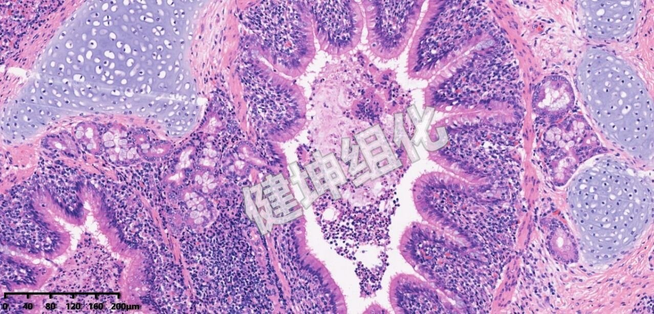
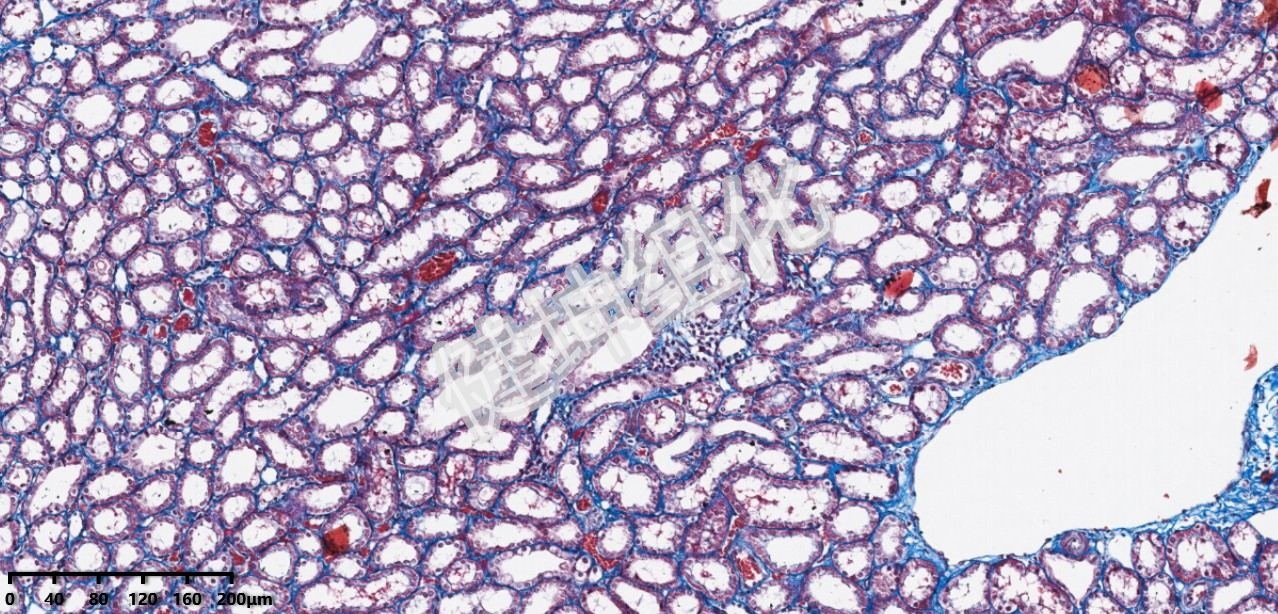
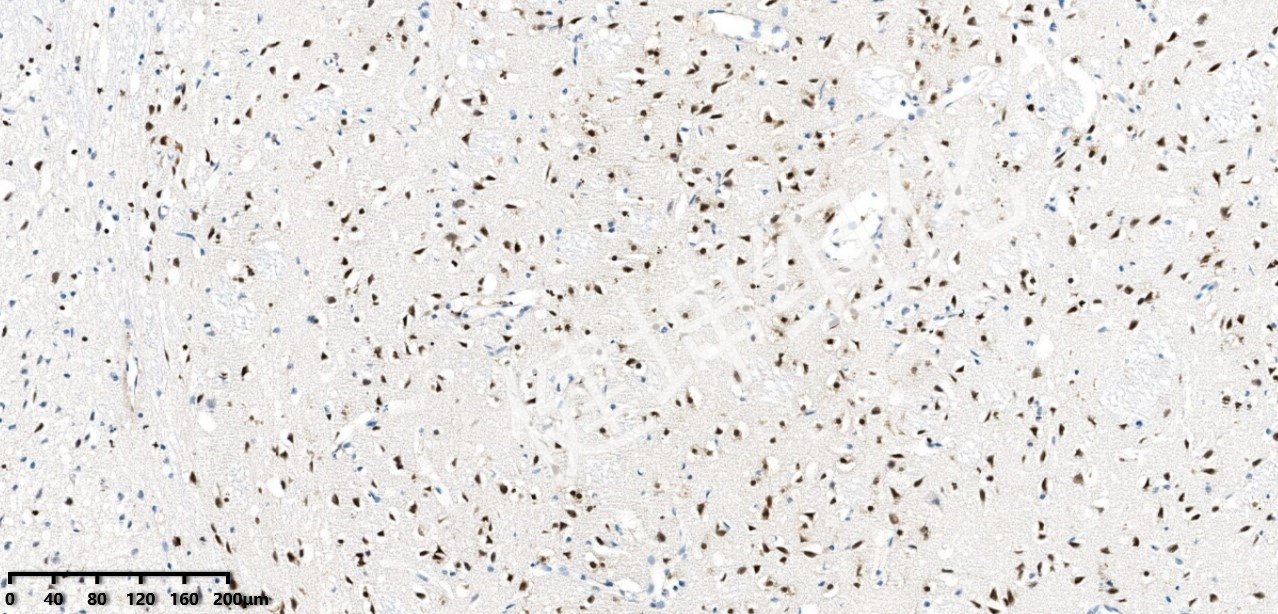

Standardized staining and pathology QC make every slide clear, reliable, and verifiable
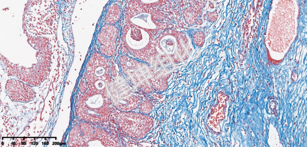
From pathological changes and organ toxicity to tissue-injury scoring, we deliver research outputs that pair specimen images with evaluation results.
Systematic observation and assessment of changes in tissue architecture and cellular morphology.
Integrated with specimen slides and experimental context to support assessment of organ-toxicity-related findings.
Presented through a standardized scoring system, facilitating statistical analysis and comparison.
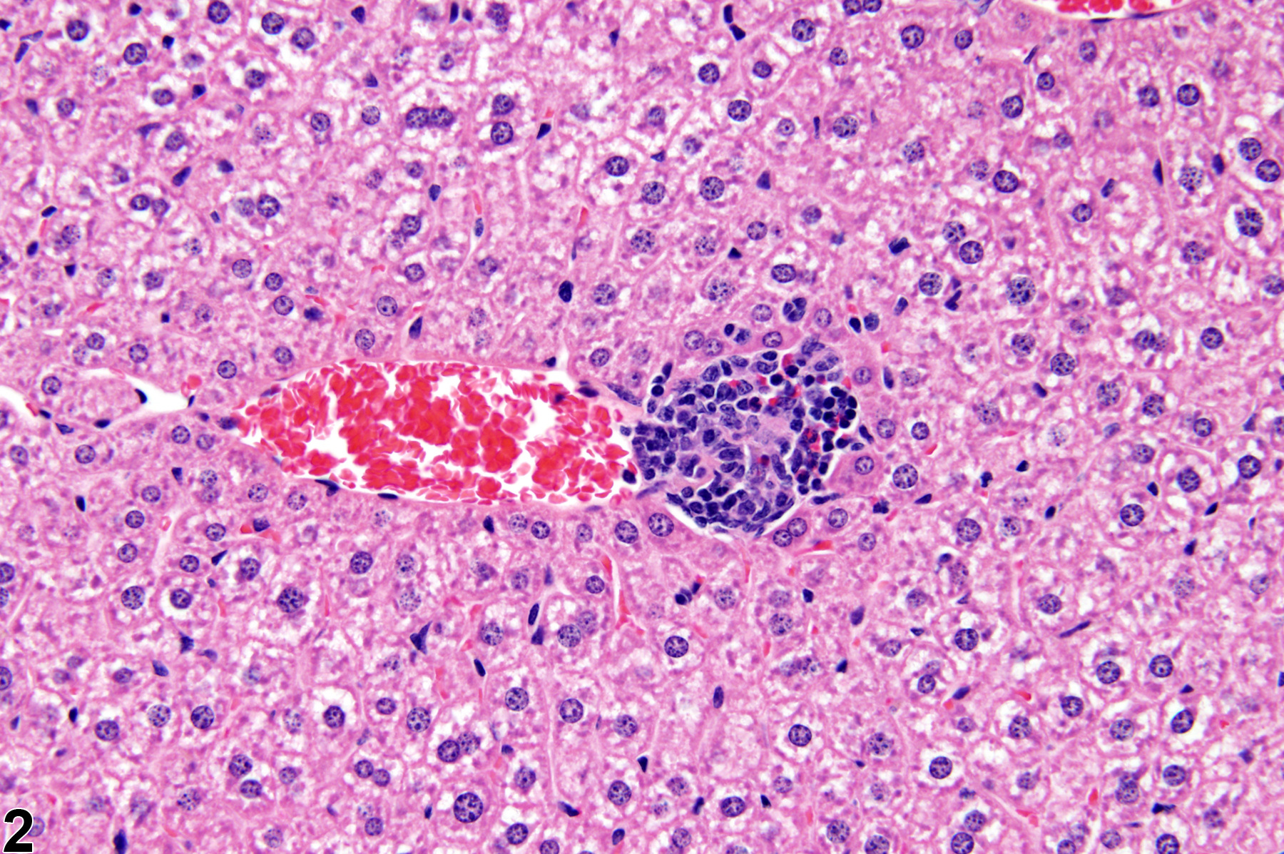
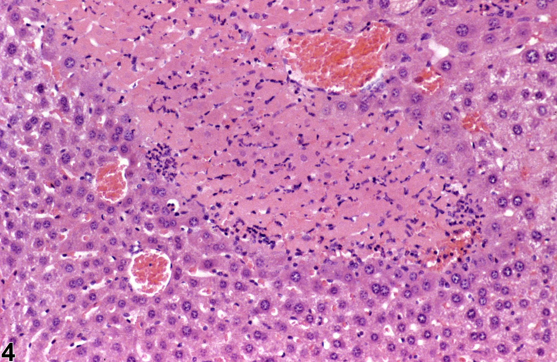
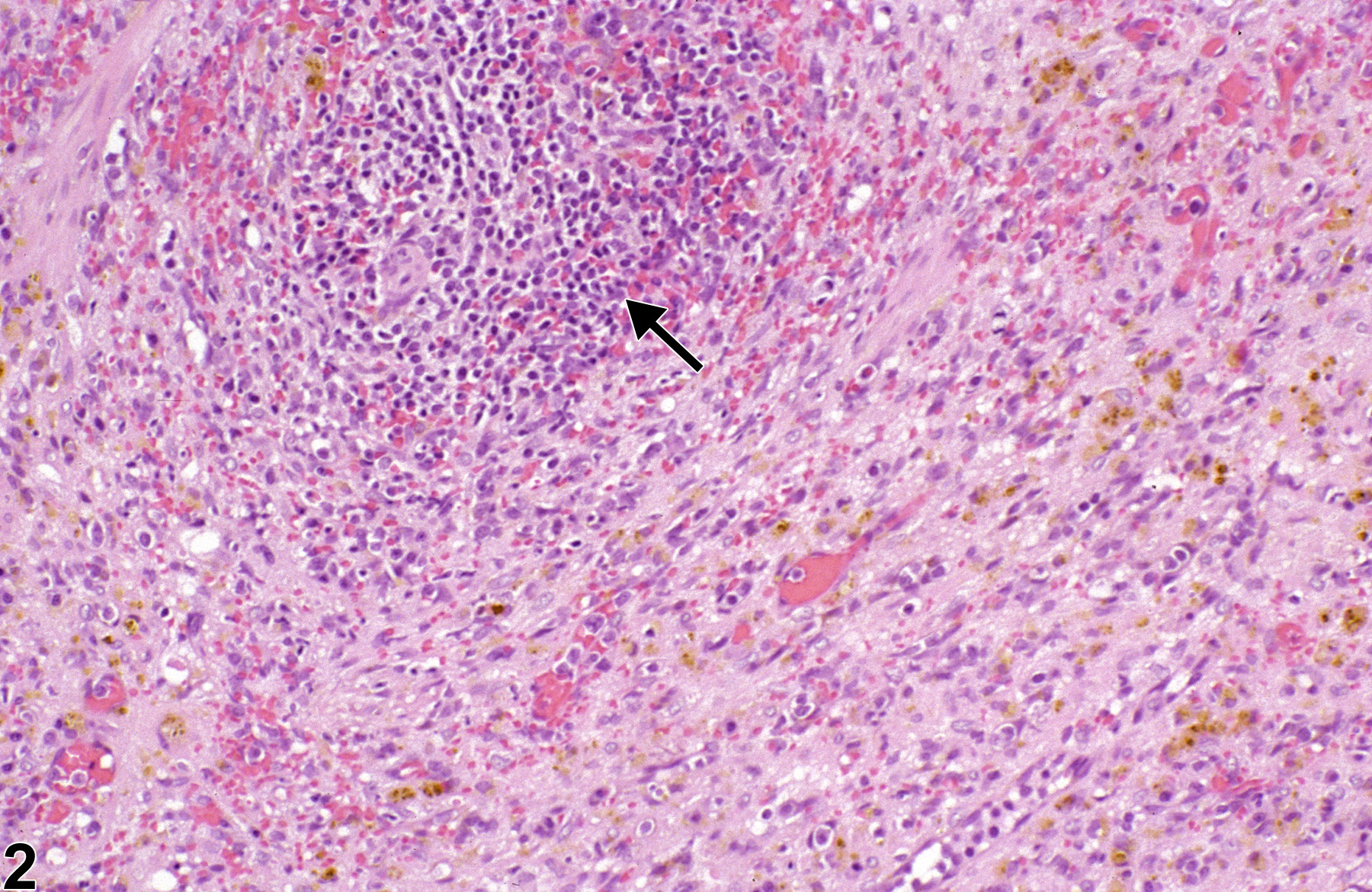
A stable immunohistochemistry and immunofluorescence system built around antibody, background, signal, localization, and reproducibility
A mature antibody-validation framework ensures experimental success rates and marker specificity that meet research standards.
Precise control of non-specific staining improves the signal-to-noise ratio, keeping images clean and reliable.
High inter-batch consistency with clear, reproducible positive signals provides stable data for research analysis.
Optimized antigen retrieval and staining conditions achieve accurate target-protein localization, making scientific results intuitive and credible.
Standardized workflows and operating procedures ensure reproducible experiments that support long-term research programs.
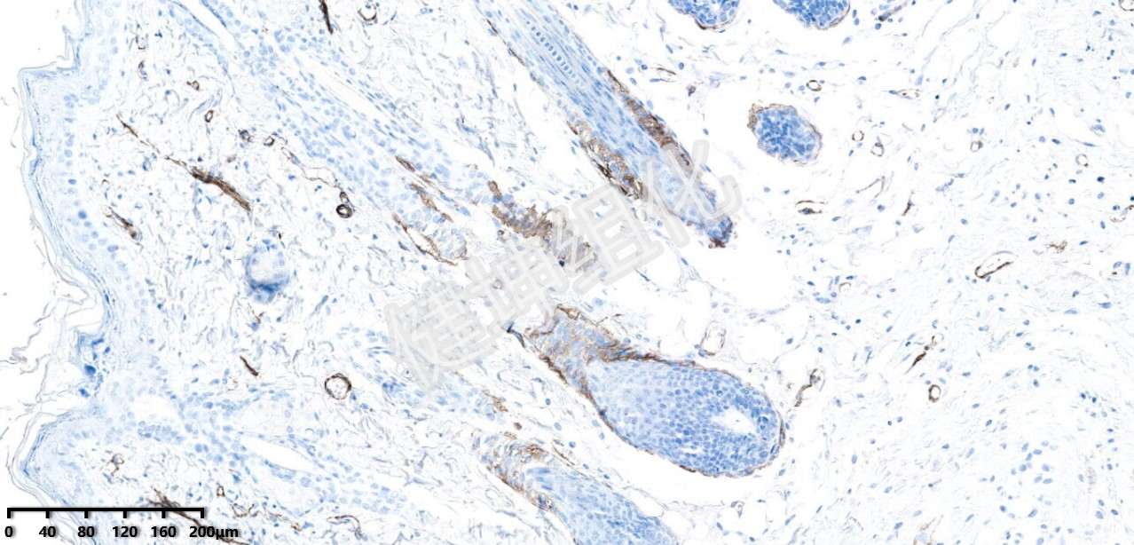
Precisely reveal target localization and expression differences, providing clear, reliable, and verifiable pathology evidence for mechanistic research, efficacy evaluation, and clinical development.
Taking expression results from "visible" to "quantifiable, statistically analyzable, and publishable"
Quantifies positive-expression intensity using metrics such as AOD and IOD—a widely used approach in immunohistochemistry research.
Quantifies the proportion of positive regions within the tissue area to assess changes in target-protein expression range.
Statistical analysis of positive-cell counts, suitable for inflammation, oncology, and immunology studies.
From histological section preparation and staining, through immunohistochemistry, multiplex immunohistochemistry, digital scanning, and quantitative analysis, every slide, every stain, and every dataset ultimately serves the advancement of research projects, the progress of studies, and the output of academic results.
Contact a project consultant